MENINGIOMA
What is a Meningioma?
The Meningioma tumor arises from the meninges – the membranes that surround your brain and spinal cord. Some meningiomas and small developing ones sometimes will not have any symptoms associated with them. Symptoms depend on the location and occur as a result of pressing the tumor on nearby tissues. Occasionally there may be headaches, vomiting, convulsions, dementia, speech problems, vision problems, unilateral weakness, or loss of bladder control. They will often become quite large before diagnosis.
Although they are usually benign (not cancerous), they can still be as big as life-threatening. Some patients may have multiple meningiomas. Some meningiomas contain cyst or calcified minerals and others contain hundreds of tiny blood vessels. As meningiomas grow inward, they usually put pressure on the brain or spinal cord. They can also grow on the outside, making the skull thicker (hyperostosis). They seem to be able to form from a variety of cells, including arachnoid cells.
How are Meningiomas Diagnosed?
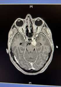
As the most common type of brain tumor in adults, most are found because of the symptoms they cause. Usually, a person will have headaches, nausea, or pain which lead to additional testing. Then if they have one, it can be confirmed on an MRI and/or a CT scan. The brain MRI scan allows precise localization of the tumor and visualization of the anatomy of the tumor and its relation to brain structures. The CT scan of the head is less precise but it gives better details of the bone changes and bone relations to the tumor. Occasionally an angiogram is necessary for a finer understanding of the vascular system and the tumor.
A Meningioma diagnosis will also confirm the location of the tumor. They can be located anywhere in the brain or spinal cord. It is primarily a central nervous system tumor, sometimes called a CNS tumor. They can be superficial when originating at the convexity or deeper and hidden under the brain, in the meninges. Often they are located at the skull base. Normally, meningiomas are more common in women between the ages of 40 and 70 years. These are found in about 3 percent of people over the age of 60.
Meningioma Treatment Options
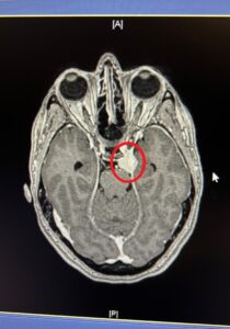 Treatment of Meningiomas is individualized for each patient based on age, size, symptoms, and location of the tumor. It may include surgical resection, radiation therapy, and observation with the aid of an MRI or a combination of the above. Complete removal of the tumor enhances the chances of the tumor not recurring. Notice the picture to the right, it’s the same MRI picture as above with the tumor circled.
- Age: One thing your doctor will ask you is if you had a previous MRI. The reason this is important is to establish the age of the tumor. Knowing how old the tumor is will help your doctor determine how fast it is growing.
- Size: The size of the tumor is important to decide if it is pushing on your brain and of course which part it’s pushing on.
- Symptoms: The symptoms you are having will be determined by where the tumor is and how big it is. As we know, different parts of the brain and spine are connected with different parts of your body. So Identifying where the tumor is, should also correlate with your symptoms.
- Location: As mentioned before, the location will help determine how important it is to have the tumor removed. If it’s in a place where it’s not affecting anything (symptoms) and not a very big size, you could possibly watch it to see if it grows bigger. Not all tumors grow. Your doctor can help you decide which option of treatment is best for you.
Grading or Classification of Tumors
After your tumor is removed, it will be sent to the lab for a grade. These grades determine the differences in the tumor. There are three types of tumors, grade i, ii, or iii. These grades are based on what the tumor looks like under a microscope at the lab that is testing it. The three types are listed below:
- Grade i tumors are the most common, 85-90 percent. The tumor is noncancerous and grows slowly. This type is also called benign tumors and is most likely not to reoccur.
- Grade ii tumors are atypical meningiomas. Around 15 to 20 percent of diagnosed tumors are grade II. Grade II’s are neither benign nor malignant (cancerous). But it can sometimes become cancerous. This type also tends to grow faster and recur.
- Grade iii are malignant or anaplastic. This is an aggressive type of meningioma. It tends to invade the closest brain tissue. Only approximately 1 to 4 percent of tumors are labeled grade III.
As you would expect, risk factors include exposure to ionizing radiation such as: during radiation therapy, family history of the condition, and neurofibromatosis type 2. As of 2014, they do not seem to be related to cell phone use. Some meningiomas contain cyst or calcified minerals and others contain hundreds of tiny blood vessels. As meningiomas grow inward, they usually put pressure on the brain or spinal cord. They can also grow on the outside, making the skull thicker (hyperostosis). They seem to be able to form from a variety of cells, including arachnoid cells.
Learn More about your Tumor
If you or a family member have been diagnosed with a Meningioma tumor, we encourage you to learn more about your tumor on our website. Your MRI radiology report generally will describe the location and type of the tumor. You can search and find the specific type of tumor. You will learn more about the nature of your type of Meningioma and how tumors of the same type and location are commonly managed at the Meningioma Center. For your convenience, feel free to send us your MRI report and scan, we will let you know the precise type and location of your tumor.
The Different Types of Meningiomas: